Detection and localization of anatomical structures in retinal images based on computer vision techniques, relational knowledge and geometric properties
Advisor: Dr.Enrique J. Carmona Suárez
In this thesis we investigate the application of a new methodology in order to perform the detection and localization of the most important anatomical structures in retinal images, such as the optic disc, the optic cup, the macula/fovea and the network of blood vessels. We address this task by means of a methodology that combine several strategies: (a) use of methods that are typically applied in this domain; (b) use of a progressive segmentation approach based on "first detection and localization after", and (c) use of relational knowledge and other characteristic properties to facilitate the approximate localization of all target structures, the exact segmentation of each of them and the detection of eye injury. This methodology is implemented using three stages: (1)In the first stage, detection of anatomical structures, several computer vision techniques are applied in order to identify typical features based on the intensity level of each pixel or his neighborhood. As a result, for each anatomical structure, we obtain a set of potential blobs. Finally, the blob most likely of belonging to each structure is chosen. This selection is based on knowledge of geometric relations between the different anatomical structures. (2)In the second stage, localization of anatomical structures, we repeat the scheme of the first stage, but focusing now in the properties and relationships between the different elements which characterize each specific anatomical structure. The rough localization of each structure obtained in the first stage allows us to simplify the processing in this stage. (3) The third stage is dedicated to the detection of ocular lesions. This stage will be addressed in a future work and will not be described in this presentation.
-
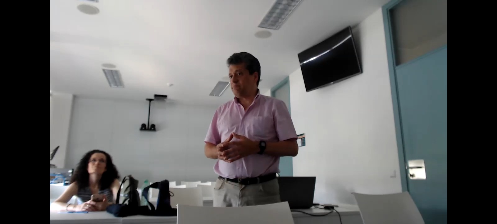
José María Molina Casado


